
Michael Zuber博士
语言
WEB资源
RESEARCH PROGRAMS AND AFFILIATIONS
研究兴趣
教育
研究抽象
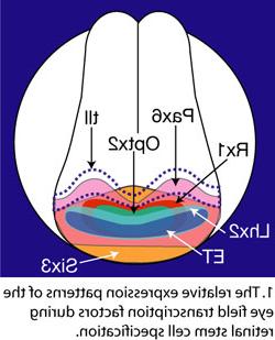
仅在美国, 10 million people are blind or suffer visual impairment due to glaucoma, 色素性视网膜炎, age-related macular degeneration and diabetic retinopathies. Every year an additional 230,000 patients loose their vision. These diseases are all due to the loss of one or more retinal cell type. Stem cells have the ability to differentiate into specialized cells. 视网膜干细胞(RS), 例如, may differentiate into a rod cell, 视网膜神经节细胞, or any of the five other cell types of the mature retina. Retinal stem cells are also self-renewing. These two characteristics make RS cells ideal for use in cell replacement therapies. Our laboratory studies the molecular mechanisms driving the specification, proliferation and differentiation of retinal stem and progenitor cells. How are Retinal Stem Cells Specified? The eye begins as a patch of ectodermal cells that are first patterned to form the beginnings of the anterior brain. These cells are further specified to become the retinal stem cells of the embryonic eye field. We have been investigating the genes responsible for retinal stem cell formation. We discovered that seven eye field transcription factors (EFTFs) are coordinately expressed in the anterior neural plate during retinal stem cell specification (Figure 1). Since each of these genes are required for normal eye formation, we reasoned that they might act together to specify retinal stem cells. 为了验证这个假设, we coexpressed all seven genes at ectopic locations in the developing frog embryo. 值得注意的是, these genes were able to induce 第三只眼睛, demonstrating that they are not only expressed during, but also sufficient for eye formation (Figure 2). Is every one of the eye field transcription factors required to generate the third eye? How do these genes interact to generate an eye? What are the downstream targets of these transcription factors? What upstream regulators coordinate the expression of these genes during eye formation? These are just a few of the questions we intend to answer in this project.

Selected Peer Reviewed Articles
Viczian AS, Bang AG, 哈里斯 WA, Zuber我. 2006. Expression of Xenopus laevis Lhx2 during eye development and evidence for divergent expression among vertebrates. Developmental Dynamics 235: 1133-1141.
Zuber我, Gestri G, Viczian AS, Barsacchi G, 哈里斯 WA. 2003. Specification of the vertebrate eye by a network of eye field transcription factors. 发展130:5155 - 5167.
Zuber我, Perron M, Philpott A, Bang A, 哈里斯 WA. 1999. Giant eyes in Xenopus laevis by overexpression of XOptx2. 细胞98:341 - 352.
Viczian AS, Vignali R, Zuber我, Barsacchi G, 哈里斯 WA. 2003. XOtx5b and XOtx2 regulate photoreceptor and bipolar fates in the Xenopus retina. 发展130:1281 - 1294.
本章
Zuber我 & 哈里斯 WA (2006). Formation of the Eye Field (pp. 8-29), in 视网膜的发展 E. Sernagor,年代. Eglen W. 哈里斯 & R. 黄(Eds.). Cambridge, UK: Cambridge University Press.